We will open our clinic all national holidays.
On november 23rd (Sat) , we will open until 19:00 pm although a national holiday.
Doctor’s schedule
We can schedule cataract surgery within 3 weeks.

What are the common problems associated with the cornea?

Dry Eye Syndrome

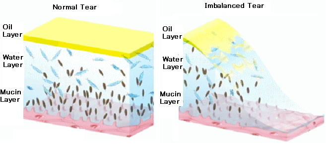
Dry eye syndrome is a chronic lack of sufficient lubrication and moisture on the surface of the eye. You can experience dry eyes when eyes don’t make enough tears or tears with the optimal chemical composition of the three layers of the eye, which are:

The outer oily layer The middle watery layer The mucin layer

Three of the layers are all crucial to lubricating, moistening and protecting the eye. If one of these 3 layers are deficient, it is referred to as "tear dysfunction syndrome", which is essentially dry eye syndrome.


Symptoms

Persistent dryness, scratching and burning in your eyes are signs of dry eye syndrome. These symptoms alone may prompt your ophthalmologist to diagnose dry eye syndrome.
But sometimes your eye doctor may want to measure the amount of tears in your eyes. A thin strip of filter paper placed under the lower eyelid, called a Schirmer test, is one way to measure tear production.
Another symptom of dry eyes is a "foreign body sensation," the feeling that something is in the eye.
And it may seem odd, but sometimes watery eyes can result from dry eye syndrome, because the excessive dryness works to overstimulate production of the watery component of your eye’s tears.

Treatments

Though dry eyes cannot be cured, there are a number of steps that can be taken to treat them. You should discuss treatment options with an ophthalmologist. Treatments for dry eyes may include:

1 Artificial tear drops and ointments.
The use of artificial teardrops is the primary treatment for dry eye. Artificial teardrops are available over the counter. No one drop works for everyone, so you might have to experiment to find the drop that works for you. If you have chronic dry eye, it is important to use the drops even when your eyes feel fine, to keep them lubricated. If your eyes dry out while you sleep, you can use a thicker lubricant, such as an ointment, at night.

2 Temporary punctal occlusion.
Sometimes it is necessary to close the ducts that drain tears out of the eye. This is first done via a painless test where a plug that will dissolve over a few days is inserted into the tear drain of the lower eyelid to determine whether permanent plugs can provide an adequate supply of tears.

3 Permanent punctal occlusion.
If temporary plugging of the tear drains works well, then silicone plugs (punctal occlusion) may be used. The plugs will hold tears around the eyes as long as they are in place. They can be removed. Rarely, the plugs may come out spontaneously or migrate down the tear drain. Many patients find that the plugs improve comfort and reduce the need for artificial tears.

4 Other medications.
Other medications, including topical steroids, may also be beneficial in some cases.

5 Surgery.
If needed, the ducts that drain tears into the nose can be permanently closed to allow more tears to remain around the eye. This is done with local anesthetic on an outpatient basis. There are no limitations in activity after having this surgery.

Keratoconus

Keratoconus is a progressive eye disease in which the normally round cornea thins and begins to bulge into a cone-like shape. This cone shape deflects light as it enters the eye on its way to the light-sensitive retina, causing distorted vision. Keratoconus can occur in one or both eyes and often begins during a person’s teens or early 20s.

Keratoconus
Picture of Keratoconus

Signs

1 Nearsightedness
2 Astigmatism
3 Blurred vision – even when wearing glasses and contact lenses
4 Glare at night
5 Light sensitivity
6 Frequent prescription changes in glasses and contact lenses
7 Eye rubbing

The first line of treatment for patients with keratoconus is to fit rigid gas permeable (RGP) contact lenses. Because this type of contact is not flexible, it creates a smooth, evenly shaped surface to see through. However, because of the cornea’s irregular shape, these lenses can be very challenging to fit. This process often requires a great deal of time and patience.
When vision deteriorates to the point that contact lenses no longer provide satisfactory vision, corneal transplant may be necessary to replace the diseased cornea with a healthy one.

Office hours

Weekday

11:30-13:45/15:00-19:30

Saturday

11:00-13:45/15:00-19:00

Sunday,Holiday

11:00-18:00
(no lunchtime.)

Closed

Our clinic is closed on 12/31, 1/1, 1/2, 1/3.

Location

9th floor of Hulic Shinjuku Building,3-25-1, Shinjuku, Shinjuku-ku, Tokyo, 160-0022

TEL:03-5363-0507

  • 1-minute walk from JR Shinjuku Station's East Gate
  • 3-minute walk from Seibu-Shinjuku Station
  • 1-minute walk from Tokyo Metro Shinjuku Station
  • 2-minute walk from Tokyo Metro Shinjuku-Sanchome Station

Map

Doctor career

Director:Yasuhiro Shinkawa

(Registered Recipient of a Diploma of Ophthalmology)

Memberships

Japan Ophthalmological Society
Japanese Retina and Vitreous Society
Japanese Society of Ophthalmic Surgeons

Certification of Completion

Course of Ophthalmic PDT Study Group
Number of cataract surgery up to the present:About 4000

Career

2001 Graduate-Medical Department of Kumamoto University
2002 Department of Ophthalmology Kyoto University School of medicine
2002 Shimada Municipal Hospital
2008 Japanese Red Cross Society
2010 Kitano Hospital The Tazuke Kofukai Medical Research Institute
2014 Shinjuku-Higashiguchi Eye Clinic


Doctor:Fumiyo Hasegawa

(Registered Recipient of a Diploma of Ophthalmology)

Memberships

Japan Ophthalmological Society
Japan Ophthalmologists Associasion
Japanese Association for Strabismus and Amblyopia(JASA)

Career

1992 Graduate- Medical Department of Teikyo Univercity
2002 The head ophthalmologist at International Catholic Hospital
2020 Shinjuku-Higashiguchi Eye Clinic

Main Thesis

Sequelae of ocular trauma in schools.(Japanese)
A case of periodic upper and lower strabismus with loss of periodicity after cataract surgery(Japanese)
Quantitative analysis of eye movement during a cover test for patients with intermittent exotropia(Japanese)


We have 7 full-time service orthoptist, 2 part-time orthoptists, 1 full-time nurse and 4 part-time nurses in our clinic.
Another several ophthalmologists are working here.