February and March's Schedule
Doctor’s schedule
We can schedule cataract surgery within 3 weeks.

Disease special page

Ophthalmologic disease

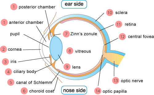
Eye Structure and Function

sp_eyeimg

associated diseases

1.posterior chamber、anterior chamber
It’s clear liquid which provides some nutrition to the cornea and the lens. It’s made from the ciliary epithelium, and it helps the eye pressure be stable.

2.cornea
It’s a clear membrane which forms the adventitia of your eye. It’s also the entrance of a ray, so it can be a lens as same as the crystalline lens.

・diffuse lamellar keratitis ( superficial punctate keratopathy)
・corneal erosion
・phlyctenular keratitis
・corneal perforation
・corneal ulcer
・corneal opacity
・herpes keratitis
・corneal dystrophy
・corneal dermoid
・bullous keratopathy
・epidemic keratoconjunctivitis
・interstitial keratitis
・keratoconjunctivitis sicca
・acanthamoeba keratitis
・keratoconjunctivitis

3.iris
It has 2 types of muscles called the iris dilator muscle and the iris sphincter muscle, and they control the amount of the light depending on the brightness.

4.ciliary body
It fixes the iris from behind and produces the aqueous fluid. It also has the ciliary muscle which changes the thickness of your lens to focus the image on your retina.

・uveitis(Harada disease)
・iridodialysis

5.canal of Schlemm
It has the ciliary vessels and the pigment particles and provides some nutrition to your retina. It also prevents from getting the unnecessary light such as a darkroom for the camera.

・uveitis(Harada disease)
・choroidal atrophy
・choroiditis

6.choroid coat
It protects the exposed part of your eyeball to make the eye movement easier.

・conjunctival concretion
・pterygium
・pinguecula
・keratoconjunctivitis sicca(dry eye)
・conjunctival foreign body
・bacterial keratoconjunctivitis
・phlyctenular conjunctivitis
・conjunctivochalasis

7.Zinn’s zonule
it’s structured by the gathering of the clear and thin fibers, and it supports the eye accommodation with your lens and ciliary body.

・ectopia Lentis

8.vitreous
it’s a clear gel which compose the most of the part of your eyeball. The light passes through it.

・posterior vitreous detachment
・vitreous hemorrhage
・synchesis scintillans
・vitreous hemorrhage
・asteroid hyalosis
・vitreous opacity
・persistent hyperplastic primary vitreous

9.lens
It’s like a lens of the camera, it focuses in conjunction with your ciliary body.

・ectopia Lentis
・lens dislocation

10.sclera
It’s a membrane which forms your eyeball, and it has a role as a body of the camera.

・episcleritis
・scleritis

11.retina
it’s like a film of the camera, and it has two photoreceptor cells which transform the light to the electric signal.

・retinal hole
・synchysis scintillans
・posterior vitreous detachment
・retinal microaneurysm
・central retinal vein occlusion
・lattice degeneration
・retinal artery occlusion

12.central fovea
It’s a part of your retina which shows you the clearest images.

・macular edema
・central retinopathy

13.optic nerve
It tells the electric signals sent from your retina to the visual cortex.

・optic neuritis
・papilledema optic neuropathy
・traumatic glaucoma
・rubeotic glaucoma
・primary open angle glaucoma
・primary angle closure glaucoma
・mixed glaucoma
・steroid induced glaucoma
・exfoliation glaucoma
・ developmental glaucoma
・malignant glaucoma
・ocular hypertension
・ optic atrophy

14.optic papilla
It’s a beginning of the optic nerve which is the bundle of the optic nerve fibers on your retina.

・ocular hypertension
・enlarged optic disc cupping
・Harada disease
・central retinal vein occlusion
・myopia
・ocular hypertension

Others

Office hours

Weekday

11:30-13:45/15:00-19:30

Saturday

11:00-13:45/15:00-19:00

Sunday,Holiday

11:00-18:00
(no lunchtime.)

Closed

Our clinic is closed on 12/31, 1/1, 1/2, 1/3.

Location

9th floor of Hulic Shinjuku Building,3-25-1, Shinjuku, Shinjuku-ku, Tokyo, 160-0022

TEL:03-5363-0507

Map

Questionnaire

SNS

TEL・FAX

Online appointment

Links

Doctor career

Director:Yasuhiro Shinkawa

(Registered Recipient of a Diploma of Ophthalmology)

Memberships

Japan Ophthalmological Society
Japanese Retina and Vitreous Society
Japanese Society of Ophthalmic Surgeons

Certification of Completion

Course of Ophthalmic PDT Study Group
Number of cataract surgery up to the present:About 4000

Career

2001 Graduate-Medical Department of Kumamoto University
2002 Department of Ophthalmology Kyoto University School of medicine
2002 Shimada Municipal Hospital
2008 Japanese Red Cross Society
2010 Kitano Hospital The Tazuke Kofukai Medical Research Institute
2014 Shinjuku-Higashiguchi Eye Clinic


Doctor:Fumiyo Hasegawa

(Registered Recipient of a Diploma of Ophthalmology)

Memberships

Japan Ophthalmological Society
Japan Ophthalmologists Associasion
Japanese Association for Strabismus and Amblyopia(JASA)

Career

1992 Graduate- Medical Department of Teikyo Univercity
2002 The head ophthalmologist at International Catholic Hospital
2020 Shinjuku-Higashiguchi Eye Clinic

Main Thesis

Sequelae of ocular trauma in schools.(Japanese)
A case of periodic upper and lower strabismus with loss of periodicity after cataract surgery(Japanese)
Quantitative analysis of eye movement during a cover test for patients with intermittent exotropia(Japanese)


We have 7 full-time service orthoptist, 2 part-time orthoptists, 1 full-time nurse and 4 part-time nurses in our clinic.
Another several ophthalmologists are working here.